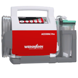
Open Use ACCUVAC or Use Suction Bag from the airway actions. Your carried equipment selects the device, with ACCUVAC taking priority when you carry both. Keep the suction tip inside the mouth and watch the fluid clear.
Start at the bottom and use short side to side sweeps toward the top. Pause, release and reassess. The illustrated path is a visual aid, not an exact target.
Sweep side to side, then release and reassess.
ACCUVAC
Take the tool. In the suction screen it starts in your hand. During laryngoscopy, select its tray slot.
Hold Left mouse and sweep. Move side to side inside the mouth. Direction changes count as a sweep; holding the tip still stops effective clearance.
Release and reassess. Confirm that the airway fluid clears and check oxygenation. Continuous suction has a separate oxygenation cost in the mod.
Need both hands for the airway? Bring the ACCUVAC tip into the mouth and press Middle mouse to park it for SALAD. It continues suction while you use the laryngoscope and tube. Press Middle mouse again to remove it. The manual bag cannot be parked.
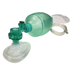
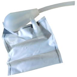
Manual suction bag
Left mouse gives one squeeze. Keep the tip in the mouth and click again after each squeeze completes. Holding the button does not repeat squeezes. Watch the collection bag fill; a full bag stops drawing.
Recognize the sound
Original mod audio. Select Play to hear each clip.
Confirm the tube’s position and reassess after moving it. A tube that advances into one main bronchus reduces the lung available for ventilation; the model reduces effective compliance and oxygenation. A partially placed obstructing tube can produce a flat capnograph.
CautionWithdrawal and sedation checks
Deflate the cuff before withdrawing the tube. Pulling repeatedly against an inflated cuff can cause airway injury. Sedation and paralysis have separate effects: rocuronium does not provide sedation or analgesia. Use the medication cards and ketamine pupil finding together.
Capnography: read the shape
A capnograph shows CO2 leaving the airway throughout each breath. Read the shape, rate and trend together with the patient’s pulse, chest movement and delivered breaths.
Normal plateau, example CO2 level 40 mmHg and breathing rate 12/min.
Adjustable teaching traces of the four game supported shapes. CO2 values and breathing rate are selected examples, not predicted patient measurements. EMMA’s numeric reading is averaged over breaths and may lag behind a change.
Normal plateau
Recognize
A quick rise, a gently rising plateau, then a sharp return toward zero.
In the game
The default shape when neither obstruction nor unresolved ventilator effort is selecting another pattern. A normal shape can still have a low or high CO2 value.
Check next
Compare the CO2 trend with breathing rate, delivered volume and perfusion. The shape alone does not establish adequate circulation or oxygenation.
Obstruction / shark fin
Recognize
A slower, slanted rise and a less distinct plateau. Increase obstruction severity to see the fin become more pronounced.
In the game
Blood or vomit obstructing the airway, or a partially placed tube, can select this pattern. A separate bronchospasm diagnosis is not required by the current waveform selector.
Check next
Reassess airway contents and tube position, use suction where indicated, and check the delivered breaths. Recheck the waveform after correcting the obstruction.
Curare cleft
Recognize
A notch interrupts the plateau as the patient makes an effort that is not coordinated with the ventilator. Increasing the example effort makes the notch deeper and wider.
In the game
Unresolved respiratory effort or coughing while the ventilator is driving breaths can create a cleft. An awake patient who is completely paralyzed cannot create a notch through awareness alone.
Check next
Reassess patient effort, ventilator coordination and the ongoing sedation and paralysis plan. A cleft is not a direct measurement of sedation; rocuronium provides paralysis without sedation or analgesia.
Flat trace
Recognize
No exhaled CO2 waveform. This example stays at zero regardless of the selected rate or peak.
In the game
An inserted but incompletely seated, obstructing ETT forces the flat shape. No breathing or no effective exhaled CO2 also removes the waveform.
Check next
Check the airway and device position, chest movement, actual delivered breaths and circulation immediately. A flat trace has more than one possible cause.
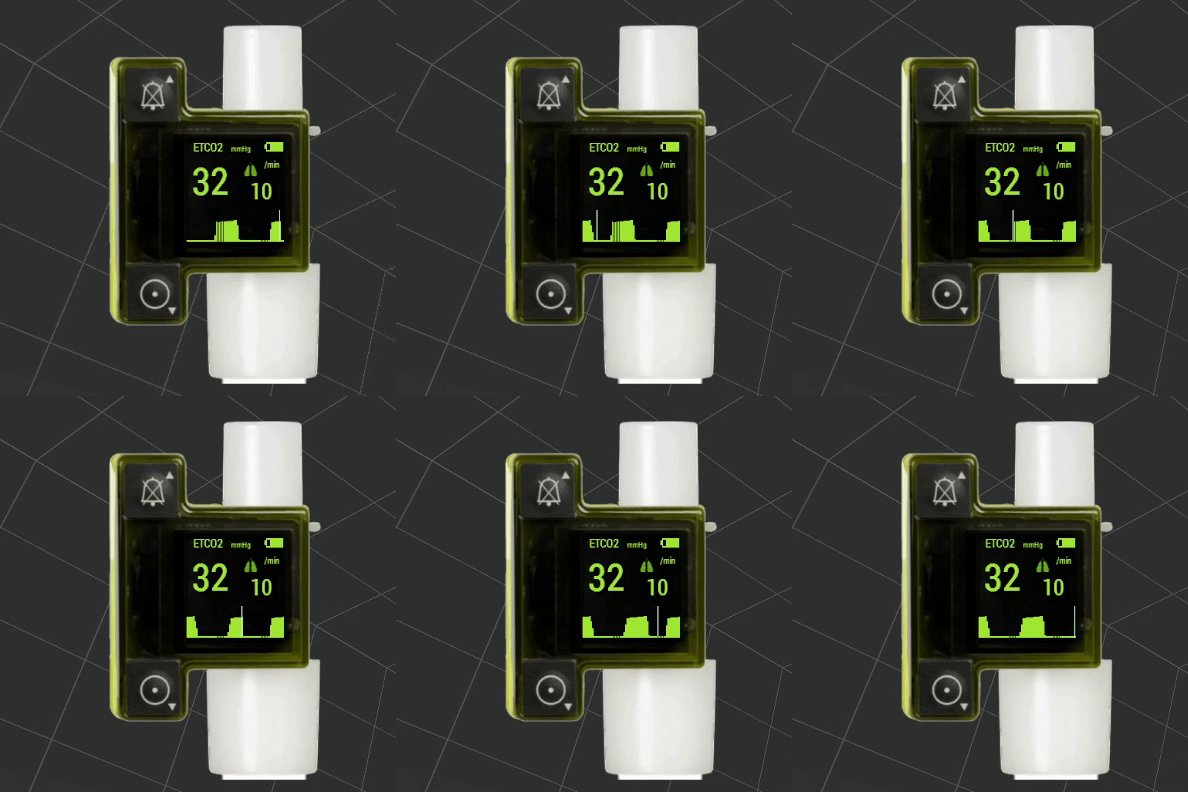
EMMA display reference
Original EMMA display reference
Waveform shape, waveform height and the breath averaged number answer different questions. Allow the number to settle after a change and reassess the patient when the shape changes.
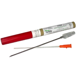
Examine the patient before placing a needle. An unnecessary decompression can create a simple pneumothorax. Continued progression depends on the air leak, available drainage, positive pressure and altitude changes.
Reassess after decompression
A decompression outlet can relieve pressure while the underlying leak remains. Reassess after placement and if the patient worsens. Repeated needles should prompt a check of the diagnosis, catheter function and need for more durable drainage.
In gameNeedle decompression option
Addon Options > ACM Extended: Trauma > Procedures > Allow Needle Decompression controls new needle placements. The procedure minigame and available equipment can add their own requirements.
Chest seals and thoracostomy
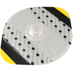
Cover the actual chest wound with the seal and inspect the other surface for additional wounds. Seal placement and peeling change the patient’s chest seal state. A seal does not remove pleural blood or clear blast lung.
Chest seal
Purpose: cover the actual open chest wound.
Recheck: seal position, other wounds and the patient’s response.
Thoracostomy
Purpose: create a pleural opening for drainage.
Recheck: opening, remaining air leak and any change after closure.
Chest tube
Purpose: maintain a drainage route, including passive pleural blood drainage.
Recheck: tube position, drainage and breathing.
Finger thoracostomy and a chest tube create different drainage conditions. The thoracostomy minigame requires site preparation, incision and completion of the opening. Inadequate preparation can seed infection. A placed tube can passively drain pleural blood, with a configured base rate of 0.12 L/min.
CautionReassess after tract closure
Closing or sealing a thoracostomy tract changes its drainage status. Reassess after closure, tube adjustment or a change in positive pressure. The chest seal tray action on a completed thoracostomy routes through the same clinical closure function as closing the tract.
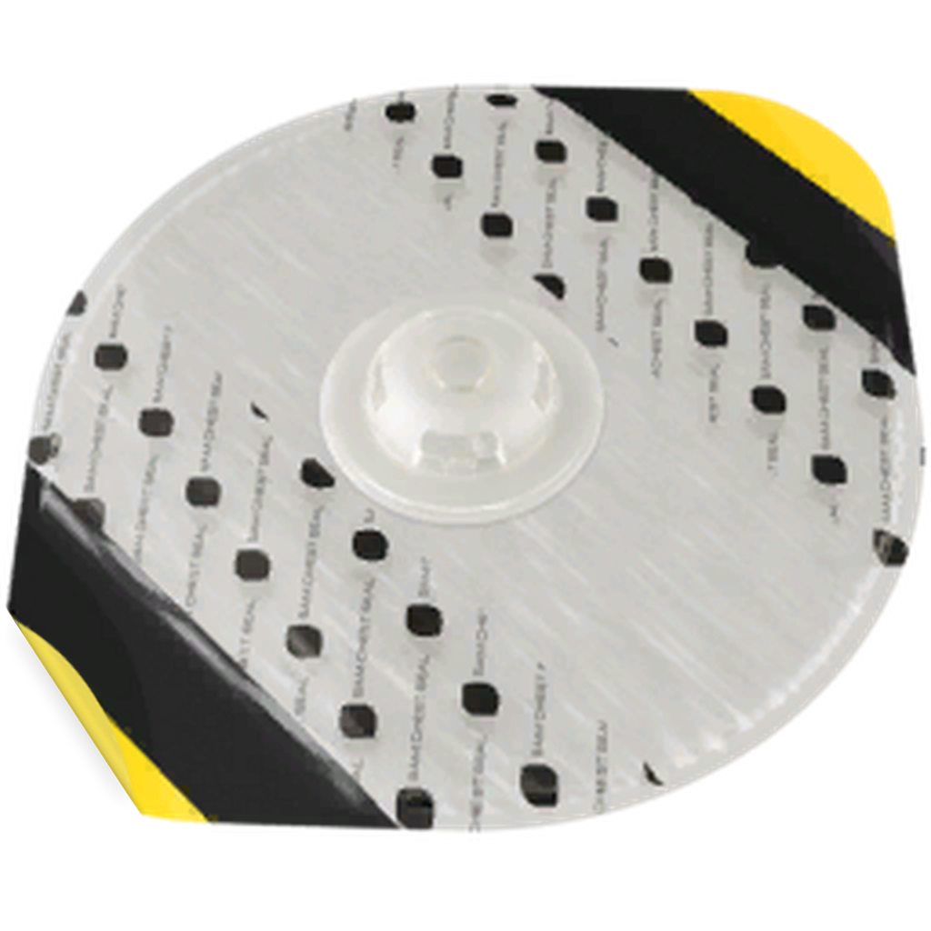
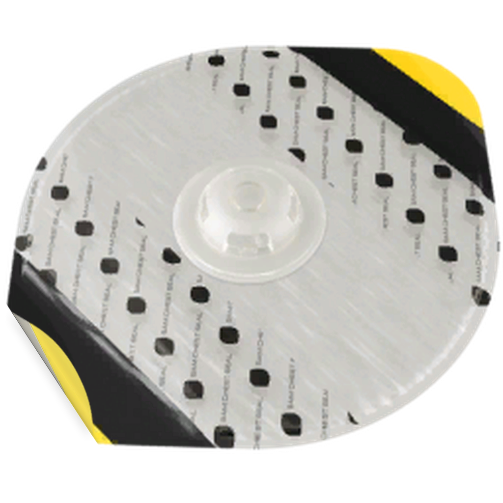
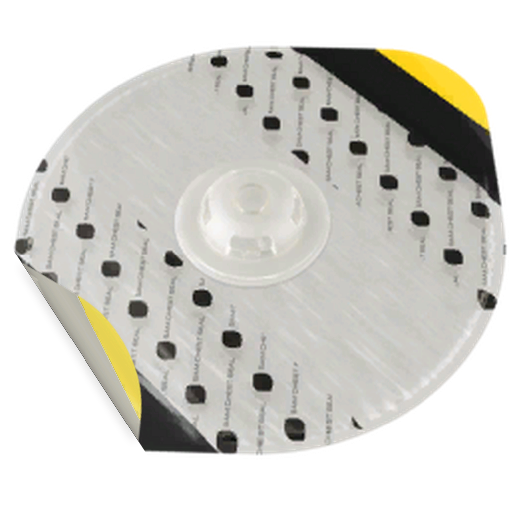
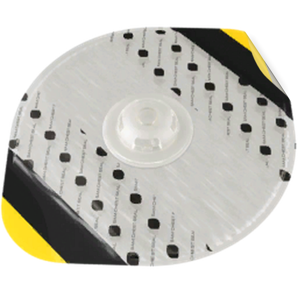
Burping a chest seal
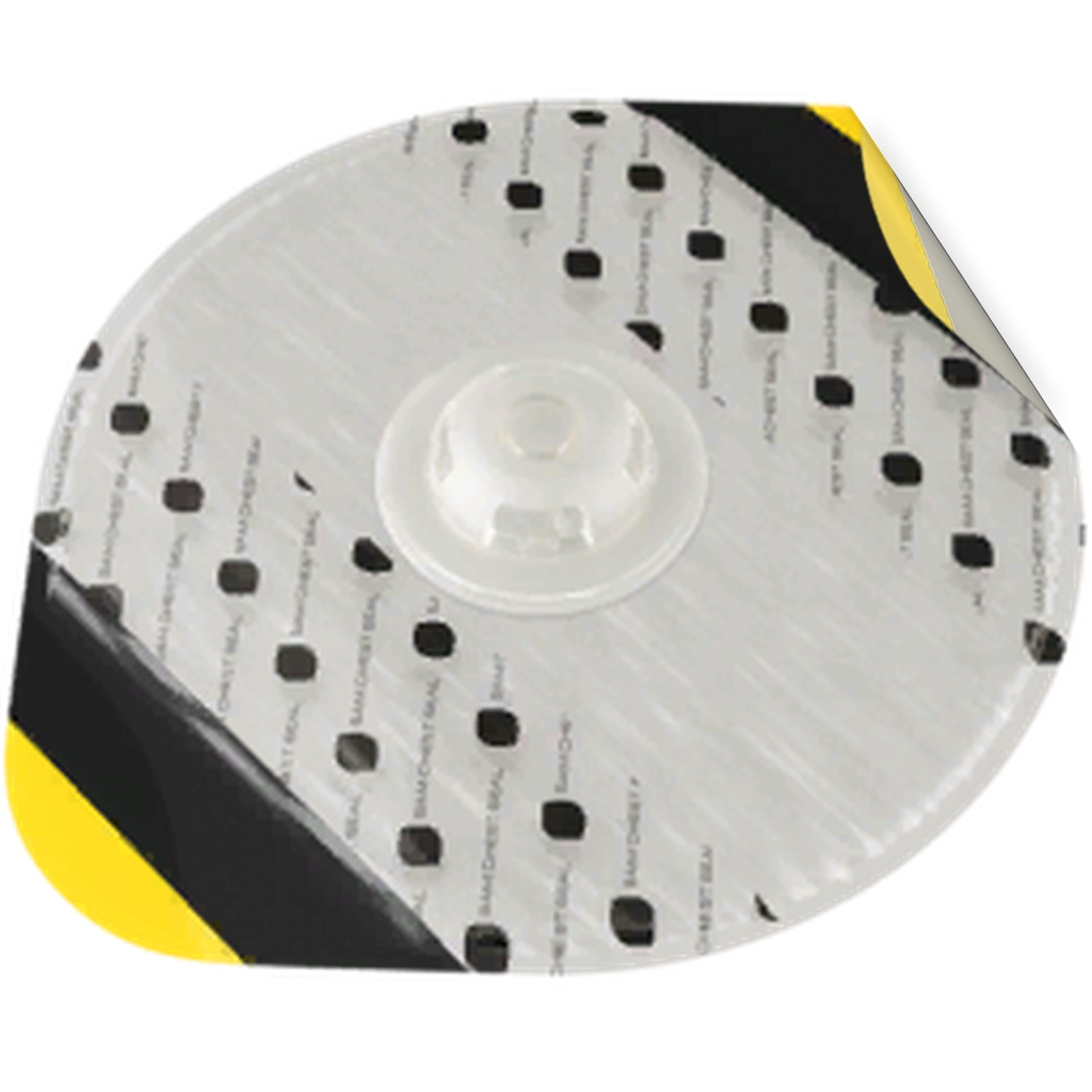
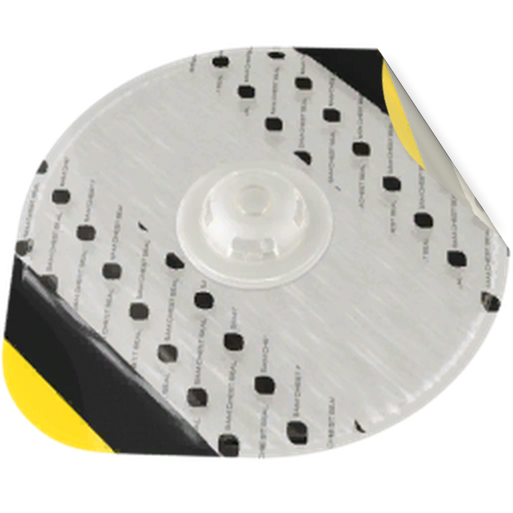
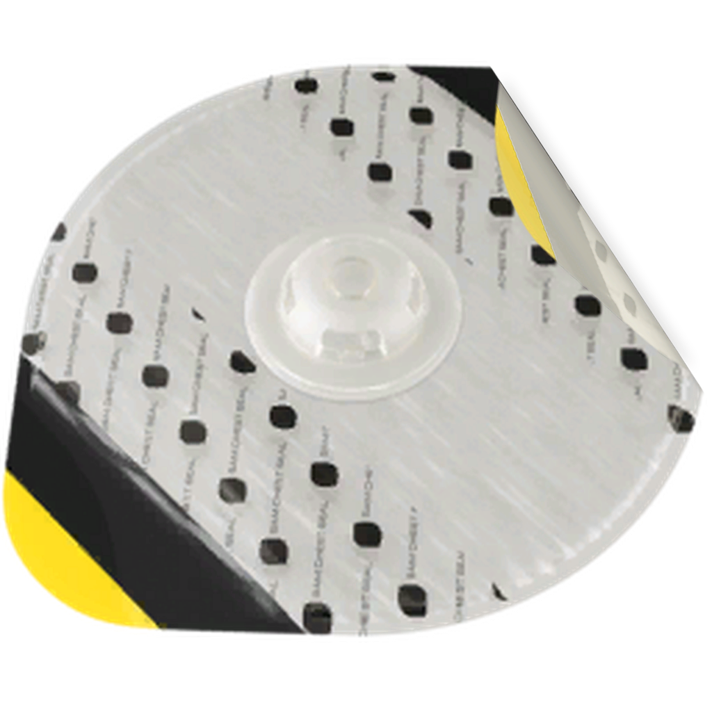
Burping lifts a corner of an applied seal to release trapped pleural air. In ACM Extended, bleeding from penetrating chest wounds can clog the seal’s outlet. If air enters faster than it escapes, the pneumothorax can grow and develop tension. Burping restores venting and gives immediate pressure relief when a seal outlet is present.
Lift the corner completely, let the seal vent, then lay it flat and reassess.
Original mod frames, eased for this demonstration. In game, each mouse wheel notch advances one frame and the seal stays where you stop. The animation timing is a viewing aid.
Use the mouse wheel
Open the chest seal screen. Put any held seal or NAR SPEAR back in its tray, then place the cursor over an applied seal.
Choose one corner. Start with Scroll down to lift the right corner, or Scroll up to lift the left. Continue in that direction for five notches to reach full lift.
Reach full lift. The burping treatment fires at the fifth frame. Stopping partway and rolling back does not trigger it. Holding at full lift does not repeatedly apply treatment.
Lay the seal back down. Reverse the wheel to lower the same corner. Moving the cursor off the seal also lays it flat and releases the corner selection.
Reassess. Check chest findings, delivered breaths, oxygenation and circulation. Check the opposite surface for a missed wound. Escalate drainage if deterioration persists or returns.
The important limit: pressure relief leaves the underlying leak and some residual collapse. A better oxygen reading after burping does not prove the pneumothorax has completely resolved.
Why it can help
Restoring an outlet lets trapped gas leave. The game reduces its pleural air burden, resets the pressure burden and clears the tension state after a completed burp with a seal outlet.
Why it can recur
Bleeding can obstruct the outlet again. An internal leak, an uncovered entry or exit wound, positive pressure or a pressure change during flight can upset the balance again.
When to recheck
Recheck after new or worsening chest findings, oxygenation or circulation, and after treatment or transport changes. A dry treated wound has no mandatory clog timer, so the mod does not require burping every seal on a fixed schedule.
The game tracks the internal air leak, accumulated pleural air, pressure and residual collapse separately. Improvement requires air entry to fall and an effective outlet to remove the trapped air. The leak can settle over time while the remaining collapse still needs drainage.
Control further air entry
Cover every communicating chest wound, including the exit. Treat ongoing bleeding. A missed opening keeps admitting air and sharply reduces the benefit of the other seals. Support breathing while avoiding unnecessary pressure on an injured lung.
Restore air drainage
A completed burp, needle decompression or an open pleural tract can relieve pressure. Keep checking the result. A chest tube provides a stronger outlet and clears the model’s residual collapse floor.
Allow the leak to settle
The internal leak gradually shrinks. Continued bleeding slows this process. Protect the patient from another chest injury, recheck after changing support, and allow drainage to keep pace while the leak settles.
What each intervention actually changes
Intervention
What can improve
What still matters
Intervention
Seal every open wound
What can improve
Reduces entry through chest wall openings and provides the seal’s venting benefit.
What still matters
Ordinary seal placement does not instantly reset established tension. Check for an obstructed outlet and retained air.
Intervention
Burp the seal fully
What can improve
Restores the seal outlet and immediately relieves the modeled pressure burden when a seal outlet is present.
What still matters
The internal leak remains. Some residual collapse can remain, and the outlet can obstruct again if bleeding continues.
Intervention
Needle decompression
What can improve
Provides a drainage outlet and relieves pressure after successful placement.
What still matters
It does not close the internal leak or remove the residual collapse floor. Reassess the patient and the actual outlet.
Intervention
Complete finger thoracostomy
What can improve
An open, completed tract creates a strong pleural outlet and relieves pressure.
What still matters
A partial incision or dissection is not yet a patent outlet. Closing or sealing the completed tract removes its open drainage route.
Intervention
Insert a chest tube
What can improve
Maintains a strong outlet, clears the residual collapse floor and allows remaining pleural air to drain toward zero.
What still matters
New air entry can still outpace the treatment. Pleural blood, edema and blast lung are separate problems that may keep breathing abnormal.
Intervention
Oxygen and ventilation
What can improve
Support oxygenation and delivered breaths while the chest problem is managed.
What still matters
Oxygen does not directly drain the pleural space or accelerate the PTX leak settling term. Actual positive pressure increases flow through an existing internal leak.
Relieved tension is not complete resolution. In the current game model, burping, a needle and an open finger tract can leave a small residual pneumothorax. Simply waiting does not remove that floor. A chest tube clears the floor so ongoing drainage can reduce the remaining air to zero. Separate full heal actions can also reset the condition.
Look for a sustained response
Breathing and delivery
Falling PTX severity reduces its penalties to lung compliance, delivered ventilation and oxygenation. Reassess chest movement, breath sounds, ventilator pressures and delivered volume together.
Circulation and oxygenation
Relieving tension removes the tension state. Continue reassessing pulse and blood pressure as well as oxygenation; hemorrhage or another injury may still be driving deterioration. SpO2 alone is unreliable in a poorly perfused patient.
Remaining chest findings
The source keeps diminished lung sounds while any PTX remains. Hemothorax, edema, blast lung or a misplaced ETT can also explain abnormal findings after the air problem improves.
How the game handles healing time
Pneumothorax leak settling time, under ACM Extended: Trauma > Pneumothorax, defaults to 600 seconds. This is a scale for reducing a normalized leak of 1 to zero, not a fixed ten minute recovery for every patient. A leak of 0.25 takes about 150 seconds with no ongoing bleeding; the same leak takes about 225 seconds at the model’s maximum bleeding modifier. New injury can increase the leak again.
The leak decreases even if the patient is unconscious or sedated. Sedation, a particular blood pressure and a particular ventilator mode are not prerequisites for this PTX healing term. Open chest wounds still admit air until covered, and actual positive pressure increases entry through an existing leak.
The default 60 second Pneumothorax stability interval only records a period of controlled air accumulation internally. It does not clear residual collapse, treat tension or produce a provider notification. Established tension still needs venting; a stopped leak and a quiet timer do not remove it.
Positive pressure can reduce estimated stroke volume when circulating volume is low. Both active BVM ventilation and the ventilator participate. Higher PEEP can increase the penalty. Treat hemorrhage and inadequate ventilation together, then reassess pressure and pulse after starting support.
The remaining airway mechanics were reviewed against ACM Extended source 4848f63. The burping and pneumothorax recovery sections use dd50edf; capnography and suction use the revisions linked in their sections. Game defaults can change with mission settings. Waveform conditions · EMMA readout · Tube mechanics · Seal effects · Thoracostomy defaults
 Manual suction bag
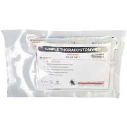 Thoracostomy
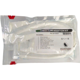 Chest tube